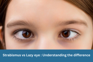
Strabismus vs Lazy eye: Understanding the difference
Blue light glasses: Do they actually work or is it marketing? Blue light glasses have become one of the most heavily marketed eyewear products in

Blue light glasses: Do they actually work or is it marketing?
Blue light glasses: Do they actually work or is it marketing? Blue light glasses have become one of the most heavily marketed eyewear products in

Smoking and your eyes: The serious risks most people do not know about
Toric IOLs for astigmatism: Do they really reduce the need for glasses after cataract surgery? When people think about the dangers of smoking, lung disease
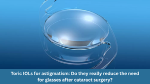
Toric IOLs for astigmatism: Do they really reduce the need for glasses after cataract surgery?
Toric IOLs for astigmatism: Do they really reduce the need for glasses after cataract surgery? Cataract surgery today is not just about removing a cloudy
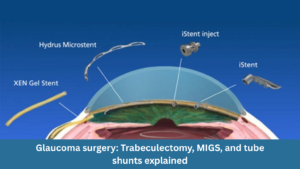
Glaucoma surgery: trabeculectomy, MIGS, and tube shunts explained
Glaucoma surgery: trabeculectomy, MIGS, and tube shunts explained Glaucoma is often called the “silent thief of sight” because it can gradually damage vision without obvious
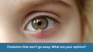
Chalazion that won’t go away: What are your options?
Chalazion that won’t go away: What are your options? A small lump on your eyelid that simply refuses to disappear can be frustrating. While many
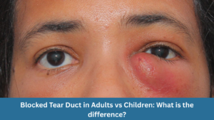
Blocked Tear Duct in Adults vs Children: What is the difference?
Blocked Tear Duct in Adults vs Children: What is the difference? A blocked tear duct, also known as nasolacrimal duct obstruction, is a common eye
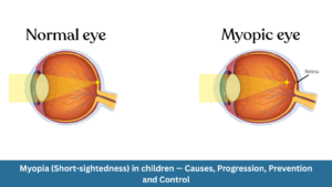
Myopia (Short-sightedness) in Children — Causes, Progression, Prevention and Control
Myopia (Short-sightedness) in Children — Causes, Progression, Prevention and Control Myopia, commonly known as short-sightedness, is becoming increasingly common among children worldwide. Many parents first
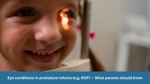
Eye conditions in premature infants (e.g. ROP) — What parents should know
Eye conditions in premature infants (e.g. ROP) — What parents should know Premature infants face a range of medical challenges, and their eyes are no
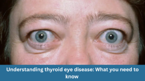
Understanding thyroid eye disease: What you need to know
Understanding thyroid eye disease: What you need to know At Clarity Eye Surgeons in Canberra, our team of skilled ophthalmologists in Canberra brings years of
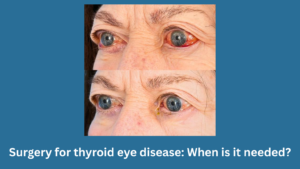
Surgery for thyroid eye disease: When is it needed?
Surgery for thyroid eye disease: When is it needed? At Clarity Eye Surgeons in Canberra, our team of experienced ophthalmologists, led by Dr Parth Shah,
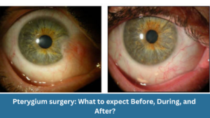
Pterygium surgery: What to expect before, during, and after?
Pterygium surgery: What to expect before, during, and after? At Clarity Eye Surgeons in Canberra, patients receive expert care led by Dr Parth Shah, a
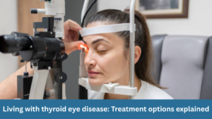
Living with thyroid eye disease: Treatment options explained
Living with thyroid eye disease: Treatment options explained At Clarity Eye Surgeons in Canberra, our team of highly qualified ophthalmologists has extensive experience diagnosing and
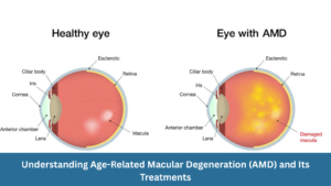
Understanding Age-Related Macular Degeneration (AMD) and Its Treatments
Understanding Age-Related Macular Degeneration (AMD) and Its Treatments At Clarity Eye Surgeons, we bring decades of ophthalmic expertise and advanced surgical experience to help Canberra
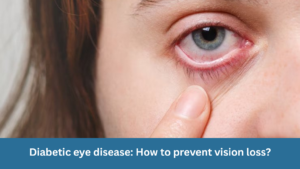
Diabetic eye disease: How to prevent vision loss?
Diabetic eye disease: How to prevent vision loss? What is diabetic eye disease? Diabetic eye disease refers to a group of eye conditions that can
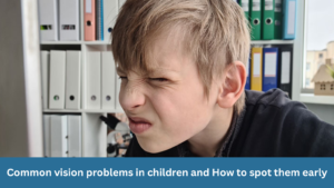
Common vision problems in children and how to spot them early
Common vision problems in children and how to spot them early At Clarity Eye Surgeons in Canberra, our team of highly experienced paediatric ophthalmologists and

Double vision in children: What parents should know
Double vision in children: What parents should know Double vision, medically called diplopia, occurs when a child sees two images of a single object instead

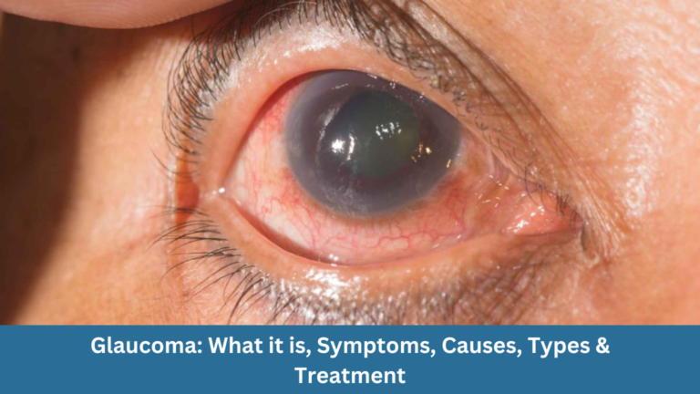
Glaucoma: What it is, Symptoms, Causes, Types & Treatment
Glaucoma: What it is, Symptoms, Causes, Types & Treatment Glaucoma is often called the “silent thief of sight” because it can damage your vision gradually,

Living with wet macular degeneration: Treatment options and lifestyle tips
Living with wet macular degeneration: Treatment options and lifestyle tips Wet macular degeneration, also known as neovascular age-related macular degeneration (AMD), is a chronic eye

Retinal detachment: Early signs you should never ignore
Retinal detachment: Early signs you should never ignore Your eyes are extraordinary organs that allow you to experience the world in vivid colour, detail, and

Vitrectomy: What you need to know
Vitrectomy: What you need to know Vitrectomy is a specialised eye surgery performed to treat a range of disorders affecting the retina and vitreous —

Diabetes and your eyes: What you need to know
Diabetes and your eyes: What you need to know Diabetes is a chronic condition that affects how your body regulates blood sugar (glucose). While most

Nystagmus: Symptoms, Causes, and Treatments
Nystagmus: Symptoms, Causes, and Treatments Nystagmus is a condition in which the eyes make uncontrolled, repetitive movements. These movements can be slow or fast, side-to-side
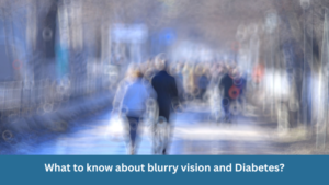
What to know about blurry vision and diabetes?
What to know about blurry vision and diabetes? Blurry vision is a common and often early symptom of diabetes. If you or someone you know
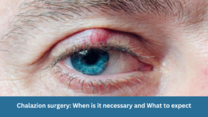
Chalazion surgery: When is it necessary and What to expect
Chalazion surgery: When is it necessary and What to expect A chalazion is a common eyelid condition that can lead to discomfort, swelling, and cosmetic
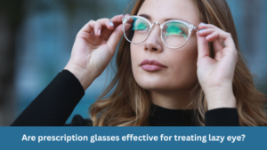
Are prescription glasses effective for treating lazy eye?
Are prescription glasses effective for treating lazy eye? Lazy eye, also known as amblyopia, is a common vision problem in children, affecting a small percentage

Dry Eye Syndrome: Symptoms, Causes & Treatment
Table of Contents Toggle What is Retinal Detachment? Causes of Retinal Detachment Symptoms of Retinal Detachment Diagnosis of retinal detachment Treatment Options for Retinal Detachment
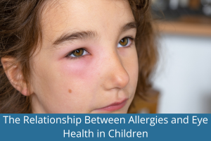
The Relationship Between Allergies and Eye Health in Children
Table of Contents Toggle 1. Symptoms of Allergies Affecting Eye Health in Children 2. Causes of Allergies Affecting Eye Health in Children 3. Potential Complications
Understanding Astigmatism: Symptoms, Causes, Diagnosis, Treatment Options & Prevention
Table of Contents Toggle What is Astigmatism? Sign & Symptoms of Astigmatism Causes of Astigmatism Types of Astigmatism Diagnosis Treatment Options for Astigmatism Astigmatism in
Understanding Retinal Detachment: Causes, Symptoms, Diagnosis and Treatment
Table of Contents Toggle What is Retinal Detachment? Causes of Retinal Detachment Symptoms of Retinal Detachment Diagnosis of retinal detachment Treatment Options for Retinal Detachment