Table of Contents
ToggleUnderstanding Retinal Detachment: Causes, Symptoms, Diagnosis and Treatment
What is Retinal Detachment?
The retina is a critical layer of your eye responsible for capturing visual images and sending them to the brain as electrical impulses. Retinal detachment occurs when the retina peels away from the underlying tissue (choroid). This separation disrupts the normal functioning of the retina and, if left untreated, can result in permanent and long-term vision loss.

Causes of Retinal Detachment
Retinal detachment can occur for a number of reasons. Below are some of the common risk factors for developing a retinal tear that can lead to retinal detachment.
- Age: As we get older, the vitreous gel inside the eye may shrink and pull away from the retina, increasing the risk of retinal detachment.
- Eye Trauma: Trauma to the eye, such as a direct blow or injury, can cause retinal detachment. Even seemingly minor injuries should be evaluated by an eye specialist.
- Previous Eye Surgeries: Individuals who have undergone cataract surgery or other eye procedures may be at higher risk of retinal detachment.
- Myopia (Nearsightedness): People with severe near-sightedness are more susceptible to retinal detachment, because their eyes are longer and the tissues are more stretched, leading to a higher risk of retinal tear.
- Other Eye Conditions: Conditions like diabetic retinopathy and inflammatory eye diseases can also elevate the risk.
Symptoms of Retinal Detachment
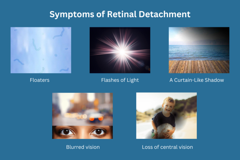
Recognising the symptoms of retinal detachment is crucial for seeking prompt medical attention. Common symptoms include:
- Floaters: Seeing a sudden onset of specks or cobweb-like shapes in your field of vision.
- Flashes of Light: Sudden bursts of light, especially in peripheral vision.
- A Curtain-Like Shadow: A shadow or dark area that seems to move across your field of vision.
- Blurred vision.
- Loss of central vision.
If you experience any of these symptoms, do not delay; contact an eye specialist immediately.
Diagnosis of retinal detachment
Diagnosing retinal detachment is a critical step in the management of this condition. Early and accurate diagnosis allows for timely intervention, which is crucial to preserving your vision. Here i’s a detailed look at the diagnostic process:
- Comprehensive Eye Examination: A thorough eye exam by an optometrist or ophthalmologist is the initial step.
- Medical History: Your medical history and risk factors are assessed.
- Visual Acuity Test: Assess central vision using an eye chart.
- Slit-Lamp Examination: Examine the front and back of the eye for abnormalities.
- Pupil Dilation: Enlarge pupils for a better view of the retina.
- Indirect Ophthalmoscopy: Use a special lens to examine the retina.
- Imaging Tests: These include ultrasound (B-scan) and optical coherence tomography (OCT).
- Differential Diagnosis: Rule out other conditions with similar symptoms.
- Prompt Referral: Confirmed cases are referred for further evaluation and treatment by a retinal surgeon. Early detection is crucial for preserving vision.
The diagnosis of retinal detachment involves a combination of a thorough eye examination, imaging tests, symptom evaluation, and, if necessary, specialised diagnostic procedures. Early detection and timely intervention are crucial to ensuring the best possible outcome for preserving your vision. If you or someone you know experiences symptoms suggestive of retinal detachment, do not delay seeking professional medical attention to assess and address the condition promptly.
Treatment Options for Retinal Detachment
Retinal detachment is a serious eye condition that demands prompt medical attention. The choice of treatment depends on various factors, including the type and extent of detachment.
Here are the primary treatment options:
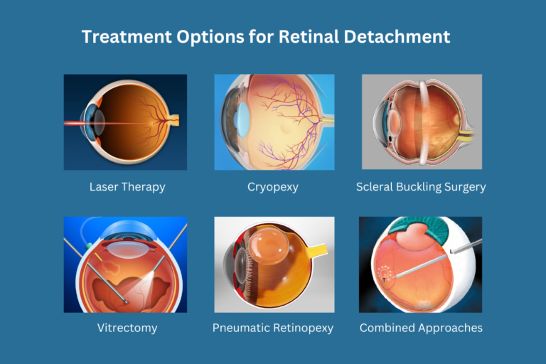
1. Laser Therapy (Photocoagulation):
- How it works: Laser therapy, or photocoagulation, is a non-invasive procedure. It involves using a laser beam to create small burns around the retinal tear. These burns form scar tissue, which seals the tear and prevents fluid from passing through and detaching the retina further.
- Indications: Laser therapy is often used for small, uncomplicated retinal tears.
2. Cryopexy (Freezing Treatment):
- How it works: Cryopexy is another method to treat retinal tears. In this procedure, a specialised freezing probe is applied externally to the eye over the tear. The extreme cold creates a scar, which holds the retina in place and prevents detachment.
- Indications: Cryopexy is suitable for specific types of retinal tears and detachments.
3. Scleral Buckling Surgery:
- How it works: Scleral buckling is a surgical procedure that involves placing a silicone band or buckle around the eye to gently push the wall of the eye inward, repositioning the detached retina against the eye wall. This technique reduces tension on the retina and allows it to reattach.
- Indications: Scleral buckling is commonly used for retinal detachments and can be effective for a variety of tear locations.
4. Vitrectomy:
- How it works: A vitrectomy is a surgical procedure that involves removing the vitreous gel from the eye, along with any scar tissue or debris that may be pulling on the retina. After this removal, a gas or silicone oil bubble is often injected into the eye to help reattach the retina. Laser is used to seal retinal breaks.
- Indications: Vitrectomy is typically used for more complex cases of retinal detachment, including those with significant scar tissue or vitreous haemorrhage.
5. Pneumatic Retinopexy:
- How it works: In this procedure, a gas bubble is injected into the vitreous cavity of the eye. The patient is then positioned in a way that allows the bubble to press against the detached retina, and a laser is then used to seal the tear.
- Indications: Pneumatic retinopexy is suitable for certain types of retinal detachments, often when the detachment is uncomplicated and can be treated without surgery.
6. Combined Approaches:
- In some cases, a combination of treatment methods may be necessary to achieve successful reattachment. For instance, laser or cryopexy may be used in conjunction with vitrectomy or scleral buckling.
- The choice of treatment or combination of treatments is determined by the ophthalmologist based on the specific characteristics of the detachment.
It is important to note that the success of retinal detachment treatment can vary depending on factors like the size, location, and duration of detachment, as well as the overall health of the eye. Early intervention and regular follow-up appointments are crucial for the best possible outcome. Patients should carefully follow their ophthalmologist’s post-operative instructions and report any changes in vision or symptoms promptly.
Recovery and Post-Treatment Care
After treatment, it is important to follow your ophthalmologist’s instructions carefully. You may need to restrict certain activities temporarily and attend regular follow-up appointments to monitor your progress. Be vigilant about any new symptoms or changes in your vision.
Prevention
While not all cases of retinal detachment can be prevented, there are steps you can take to reduce your risk:
- Regular Eye Exams: Schedule routine eye examinations, especially if you have risk factors or a family history of retinal detachment.
- Eye Protection: Wear protective eyewear during sports or activities with a risk of eye injury.
- Manage Underlying Conditions: If you have diabetes or other medical conditions affecting your eyes, work closely with your healthcare team to manage them effectively.
Conclusion
Retinal detachment is a serious eye condition that requires immediate attention and treatment. Early detection and intervention can make all the difference in preserving your vision. If you have any concerns about your eye health or experience symptoms like floaters, flashes of light, or shadows in your vision, don’’t hesitate to reach out to Clarity Eye Surgeons for expert care and guidance.
For more information or to schedule an appointment, please contact us.
Author Bio
Dr Parth Shah is a director and principal ophthalmologist at Clarity Eye Surgeons in Canberra, specialising in cataract surgery and strabismus surgery. With extensive training and experience, he is renowned for his expertise in the field. Dr Shah is dedicated not only to performing successful surgeries but also to patient education. His compassionate approach, combined with technical proficiency, has earned him the trust and gratitude of countless patients. He is a true advocate for eye health and a trusted name in the Canberra ophthalmology community.
FAQs
While it is rare for retinal detachment to happen in both eyes simultaneously, individuals who have had retinal detachment in one eye are at a higher risk of developing it in the other eye. Regular eye check-ups can help monitor both eyes, especially if you’re at higher risk.
Recovery time varies depending on the type of surgery and the severity of the detachment. Most patients can expect visual improvement within a few weeks, but full recovery may take several months. Your ophthalmologist will guide you on specific post-operative care and activity restrictions.
Yes, there is a chance of recurrence even after successful treatment. Regular follow-up visits are essential to monitor healing, and any new symptoms such as floaters or flashes of light should be reported immediately to your eye specialist.
Flying is usually not recommended immediately after retinal detachment surgery, especially if a gas bubble was used in the procedure, as changes in altitude can cause pressure changes that may affect the bubble. Your ophthalmologist will advise when it is safe to travel by air.
If left untreated, retinal detachment can lead to permanent vision loss and potentially total blindness in the affected eye. Timely intervention is crucial to prevent irreversible damage to the retina.