What Is Hypotropia?
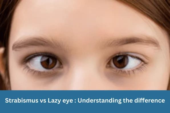
Hypotropia describes a vertical strabismus in which one eye is positioned lower than the other. Like all vertical deviations, it is described from the perspective of the lower eye — a left hypotropia means the left eye sits lower than the right. This is the exact counterpart of right hypertropia. In practice, both terms describe the same misalignment, and clinicians may use either depending on which eye is most usefully described.
The primary functional consequence is vertical diplopia— images from the two eyes are displaced vertically, making most tasks requiring both eyes to work together uncomfortable or impossible. Reading, driving, and descending stairs are particularly affected.
Causes
Orbital Floor Fracture (Blowout Fracture)
A direct blow to the orbit (from a ball, fist, or car accident) can fracture the thin floor of the eye socket. The inferior rectus muscle — which lies directly above the orbital floor — can herniate through the fracture and become entrapped. The trapped muscle cannot contract freely and restricts the eye’s upward movement.
The affected eye appears lower (hypotropic) relative to the unaffected eye because it cannot fully elevate. The patient typically presents with vertical diplopia, worst on upgaze. A trapdoor fracture variant — more common in children — can entrap the muscle without visible fracture displacement on plain imaging and may cause ischaemia of the muscle if not released promptly.
Third Nerve Palsy
The third cranial nerve innervates most of the extraocular muscles, including the superior rectus, medial rectus, inferior rectus, inferior oblique, and levator palpebrae. A complete third nerve palsy causes the eye to deviate outward and downward (from the unopposed action of the lateral rectus and superior oblique), with a complete ptosis. If the inferior rectus and inferior oblique are differentially affected, a hypotropia may be the presenting feature.
A new third nerve palsy in an adult requires urgent assessment to exclude a compressive cause such as an intracranial aneurysm, particularly when the pupil is involved.
Superior Rectus Paresis
Isolated weakness of the superior rectus (which elevates the eye) causes the affected eye to sit lower than expected, producing a hypotropia. This is relatively uncommon in isolation and may occur following orbital trauma, orbital surgery, or as part of a more generalised motility disorder.
Double Elevator Palsy
A rare condition in which both muscles responsible for elevating the eye in their primary action — the superior rectus and inferior oblique — are functionally deficient. The affected eye is deviated downward and cannot be elevated in any field of gaze. It may be congenital or acquired, and may be associated with restriction of the inferior rectus as well as paresis of the elevators.
Hypotropia as the counterpart of contralateral hypertropia
Diagnosis
The most important diagnostic distinction in hypotropia is whether the mechanism is restrictive or paretic. This determines the surgical approach and predicts the prognosis for spontaneous recovery.
Forced Duction Test
This clinical test involves passively rotating the eye under topical or sub-Tenon anaesthesia. If the eye encounters resistance when manually elevated, this indicates a restrictive cause (e.g. entrapped inferior rectus). If the eye moves freely, the problem is paretic— the muscle that should be elevating the eye is weak, not mechanically blocked.
Other key investigations include:
- • CT scan of the orbits — to identify orbital floor fracture, muscle entrapment, or soft tissue herniation
- • Hess chart — to map the full pattern of restricted and overacting muscles
- • Prism cover test in multiple gaze positions
- • Brain imaging — for new third nerve palsy without a clear cause
Experiencing vertical double vision after an eye injury?
Prompt assessment can determine whether muscle entrapment requires urgent surgical repair.
Treatment
Orbital Floor Fracture Repair
Surgical repair of the orbital floor is indicated for confirmed muscle entrapment causing significant diplopia, enophthalmos (posterior displacement of the globe), or a trapdoor fracture with evidence of muscle ischaemia. The surgeon releases the entrapped tissue and covers the bony defect with a titanium mesh or implant. If some restriction persists after fracture repair, further strabismus surgery on the muscles themselves may be required at a later stage.
Strabismus Surgery for Paretic Hypotropia
Once spontaneous recovery has been given adequate time (typically six to twelve months for traumatic or microvascular nerve palsies), surgical correction addresses the residual deviation. The appropriate procedure depends on the muscles involved. For superior rectus paresis, options include inferior rectus recession on the affected eye or a transposition procedure. For double elevator palsy, the Knapp procedure — transposing the medial and lateral rectus muscles to the superior rectus insertion — is a well-established approach.
Prism Glasses
Vertical prisms incorporated into spectacle lenses can provide useful functional relief from diplopia for small to moderate deviations. They are frequently used while awaiting surgical repair or during the observation period for nerve palsies. Fresnel press-on prisms allow rapid, non-permanent prescribing during this phase.
Timing is critical for trapdoor fractures in children
Conclusion
Hypotropia is a vertical strabismus with several distinct causes. The forced duction test is the pivotal clinical tool that differentiates restriction — which requires mechanical release — from paresis, which may recover spontaneously or require muscle surgery after an adequate waiting period. With accurate diagnosis and appropriately timed intervention, the great majority of patients achieve a significant reduction in diplopia and a functional improvement in their quality of life.
Frequently Asked Questions
They describe the same vertical misalignment from different perspectives. If the left eye is lower than the right, this is called left hypotropia (the left eye is depressed) and is exactly equivalent to right hypertropia (the right eye appears elevated). The terminology used depends on clinical convention, but the underlying condition is the same. Treatment planning focuses on the eye with the mechanical problem rather than the labelling convention.

Written by
Dr Parth ShahDr Parth Shah is a subspecialty-trained Canberra ophthalmologist with expertise in strabismus surgery, paediatric eye care and cataract surgery.
Medical disclaimer: This article is for educational purposes only and does not constitute medical advice, diagnosis, or treatment. Always consult a qualified ophthalmologist or healthcare professional regarding any eye condition or before making any healthcare decisions. Read our full disclaimer.
Ready to take the next step?
Book a consultation to assess the cause of your hypotropia and discuss the most appropriate management plan.