What Is Cyclotropia?
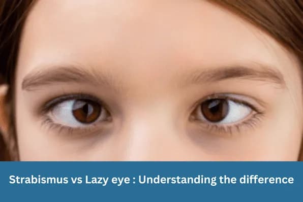
Most forms of strabismus involve eyes that deviate horizontally (inward or outward) or vertically (up or down). Cyclotropia is different — it is a torsional or rotational misalignment, where one eye is rotated around its own axis relative to the other.
The two subtypes are defined by which direction the top of the eye rotates:
- Incyclotropia— the top of the eye rotates toward the nose (intorsion).
- Excyclotropia— the top of the eye rotates away from the nose (extorsion). This is more commonly symptomatic and clinically encountered.
Cyclotropia is often associated with vertical strabismus (such as hypertropia from 4th nerve palsy) and may be part of a complex pattern of misalignment rather than an isolated finding.
Causes of Cyclotropia
The superior oblique and inferior oblique muscles are responsible for controlling the torsional position of the eye. Disruption of either muscle or its nerve supply is the most common cause of cyclotropia.
| Incyclotropia | Excyclotropia | |
|---|---|---|
| Definition | Top of eye rotated toward nose (intorsion) | Top of eye rotated away from nose (extorsion) |
| Common causes | Superior rectus palsy, iatrogenic (post-surgery) | Superior oblique palsy (4th nerve palsy), bilateral SO palsy |
| Appearance on double Maddox rod | Image tilted one way relative to the other | Image tilted the opposite way — excyclotorsion more commonly symptomatic |
| Compensatory head posture | Uncommon | Chin down, head tilt to contralateral shoulder (classic 4th nerve palsy posture) |
| Primary surgery | Superior oblique tuck (strengthens intorsion) | Inferior oblique weakening or Harada-Ito procedure |
Superior oblique palsy is the most common cause of excyclotropia in clinical practice. It may be congenital (present from birth, often with a long-standing compensatory head tilt), or acquired (from head trauma, raised intracranial pressure, or microvascular disease from hypertension or diabetes).
Skew deviation— a vertical misalignment caused by a neurological lesion in the posterior fossa or brainstem rather than an extraocular muscle problem — can produce a torsional component and must be excluded with appropriate imaging when cyclotropia develops without obvious cause.
Symptoms and Diagnosis
The hallmark symptom of cyclotropia is torsional diplopia — the two images seen (when both eyes are open) appear tilted relative to each other rather than separated horizontally or vertically. Patients often describe one image as appearing “rotated” or “diagonal.”
Other features of superior oblique palsy that frequently accompany cyclotropia include: a compensatory head tilt to the opposite shoulder, chin-down head posture, and difficulty reading or doing close work (as near tasks often worsen torsion).
Key diagnostic tool: the double Maddox rod test
Fundus photography is used objectively: the normal optic disc sits slightly above the level of the fovea; torsion shifts this relationship and can be measured from photographs.
Experiencing tilted or rotated double vision?
Torsional diplopia warrants specialist assessment. A referral from your GP is needed to see our strabismus team.
Treatment Options
Treatment decisions depend on the underlying cause, the degree of torsion, and whether spontaneous recovery is expected.
- Observation— for acute-onset cyclotropia from a microvascular cause (diabetes, hypertension), spontaneous recovery within 3–6 months is common and should be awaited before surgery is considered.
- Inferior oblique weakening— recession or myectomy of the inferior oblique muscle reduces its extorsional action and is the most common surgical approach for excyclotropia from unilateral SO palsy with moderate torsion.
- Superior oblique tuck— shortening the SO tendon to strengthen its intorsional pull. Useful when IO weakening alone is insufficient.
- Harada-Ito procedure— anteriorisation of the anterior fibres of the SO tendon, designed specifically to increase intorsion. Most effective for bilateral SO palsy with large excyclotorsion.
Surgery rarely achieves perfect torsional alignment, but a significant reduction in the degree of torsion — enough to eliminate symptomatic diplopia in primary gaze — is achievable in most cases. Adjustable suture techniques are particularly valuable for adults with cyclotropia where precise intraoperative titration is needed.
Frequently Asked Questions
They are closely related but not identical. Superior oblique (4th nerve) palsy is the most common cause of excyclotropia, but cyclotropia can also result from superior rectus palsy, skew deviation, bilateral SO palsy, or previous eye muscle surgery. Not all 4th nerve palsies cause symptoms — small torsional deviations may be well-compensated by the visual system.

Written by
Dr Parth ShahDr Parth Shah is a subspecialty-trained Canberra ophthalmologist with expertise in strabismus surgery, paediatric eye care and cataract surgery.
Medical disclaimer: This article is for educational purposes only and does not constitute medical advice, diagnosis, or treatment. Always consult a qualified ophthalmologist or healthcare professional regarding any eye condition or before making any healthcare decisions. Read our full disclaimer.
Ready to discuss your treatment options?
Our strabismus team can assess and manage cyclotropia and other complex eye muscle conditions.