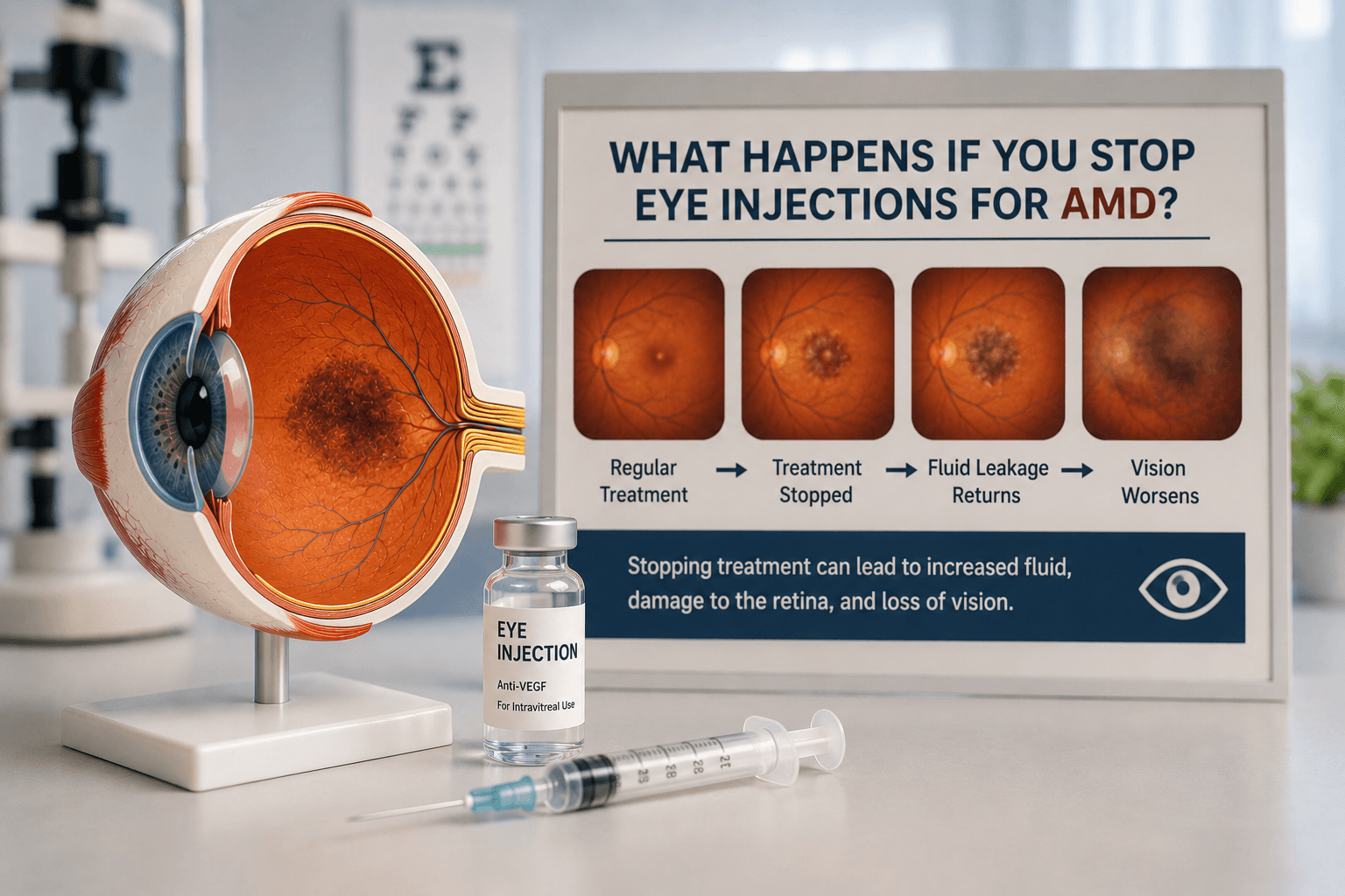
Understanding Diabetic Retinopathy
Diabetic retinopathy is damage to the blood vessels of the retina caused by chronically elevated blood glucose. It is the most common cause of new blindness in working-age Australians, and affects approximately one-third of all people with diabetes to some degree. Progression follows a broadly predictable course:
- Non-proliferative diabetic retinopathy (NPDR): The earlier stage, characterised by microaneurysms (tiny outpouchings of retinal capillaries), dot and blot haemorrhages, hard exudates (lipid deposits), and retinal oedema. NPDR ranges from mild to severe, and the severity determines the risk of progressing to the more dangerous proliferative stage.
- Proliferative diabetic retinopathy (PDR): The advanced stage, in which new, fragile blood vessels grow on the surface of the retina and into the vitreous cavity in response to retinal ischaemia. These vessels bleed easily, causing vitreous haemorrhage, and can cause tractional retinal detachment if fibrous tissue contracts.
1 in 3
People with diabetes have some degree of diabetic retinopathy
DMO
Leading cause of vision loss in working-age Australians with diabetes
PBS
Subsidises Eylea and Lucentis for diabetic macular oedema
Diabetic Macular Oedema: The Most Treatable Threat
Diabetic macular oedema (DMO) is the accumulation of fluid within the layers of the macula, caused by leakage from damaged retinal capillaries. It can occur at any stage of diabetic retinopathy — including relatively early NPDR — and is the primary mechanism by which diabetes causes central vision loss.
DMO is detected on optical coherence tomography (OCT), which allows precise measurement of retinal thickness and fluid localisation. Patients may notice blurred or distorted central vision, difficulty reading, or washed-out colour perception — though in early cases the oedema may be detectable on OCT before it causes noticeable symptoms.
Anti-VEGF Injections for DMO
VEGF (vascular endothelial growth factor) is overproduced in the ischaemic diabetic retina, driving both vascular permeability (causing DMO) and new vessel growth (causing PDR). Anti-VEGF drugs delivered directly into the vitreous cavity of the eye block this protein, reducing fluid leakage and, over time, allowing the macula to dry out.
The major anti-VEGF agents used for DMO in Australia include:
- Ranibizumab (Lucentis)— PBS-subsidised for centre-involving DMO with visual acuity impairment.
- Aflibercept (Eylea)— PBS-subsidised for centre-involving DMO; may offer slightly longer treatment intervals.
- Bevacizumab (Avastin)— used off-label; comparable efficacy to ranibizumab in large randomised trials, and significantly lower cost.
Treatment typically begins with monthly injections for 3–6 months, after which the interval is extended based on OCT response. In the landmark DRCR.net Protocol T trial, all three agents produced meaningful improvements in visual acuity at two years, with more severe DMO cases showing a larger benefit from aflibercept.
Steroid implants for refractory DMO
Anti-VEGF in Proliferative Diabetic Retinopathy
In PDR, anti-VEGF injections can cause dramatic regression of new retinal blood vessels within days of administration. This makes them valuable in several scenarios:
- • Pre-operatively before vitrectomy surgery for vitreous haemorrhage or tractional retinal detachment, to reduce surgical bleeding.
- • As a bridge when panretinal photocoagulation (PRP) laser cannot be completed in one sitting — for example, due to vitreous haemorrhage obscuring the view.
- • As an alternative to PRP in selected patients, though PRP remains the standard of care for high-risk PDR due to its durable, single-treatment effect.
The DRCR.net Protocol S trial compared anti-VEGF injections against PRP for PDR and found non-inferior visual acuity outcomes at two years for the injection arm, with the benefit of preserving peripheral visual field. However, the injection arm required ongoing treatment, emphasising the importance of patient adherence.
The Importance of Systemic Control
Eye injections treat the ocular manifestations of diabetic retinopathy but cannot substitute for good systemic diabetes management. The following are critical to reducing the burden of ocular treatment:
- Blood glucose control: Lowering HbA1c reduces the rate of retinopathy progression and the frequency of injections required.
- Blood pressure: Hypertension accelerates retinal vascular damage; target blood pressure <130/80 mmHg is recommended.
- Cholesterol: Elevated lipids contribute to hard exudate deposition and vascular damage; statin therapy is commonly indicated.
Diabetes and concerns about your vision?
Regular diabetic eye screening is essential. Book a comprehensive retinal assessment with Dr Parth Shah.
Conclusion
Anti-VEGF eye injections are a proven, effective treatment for the most vision-threatening complications of diabetic retinopathy. For DMO, they are the first-line standard of care, supported by strong trial evidence and PBS subsidy in Australia. In PDR, they complement laser and surgical approaches. The key to good outcomes is early detection through regular diabetic eye screening, prompt treatment when indicated, and good systemic diabetes management.
Frequently Asked Questions
Yes, potentially. Diabetic macular oedema (DMO) can cause significant retinal fluid on OCT imaging before it noticeably reduces your visual acuity. Treating DMO at this stage — when the retina is still in good condition — produces better final vision outcomes than waiting until acuity has already declined. Your ophthalmologist will review your OCT and visual acuity together when deciding whether treatment is warranted.

Written by
Dr Parth ShahDr Parth Shah is a subspecialty-trained Canberra ophthalmologist with expertise in cataract surgery, paediatric eye care and strabismus surgery.
Medical disclaimer: This article is for educational purposes only and does not constitute medical advice, diagnosis, or treatment. Always consult a qualified ophthalmologist or healthcare professional regarding any eye condition or before making any healthcare decisions. Read our full disclaimer.
Ready to protect your vision?
Book a consultation with Dr Parth Shah to discuss diabetic eye disease and your treatment options.