Types of Retinal Vein Occlusion
Retinal vein occlusion is the second most common retinal vascular disorder after diabetic retinopathy. It occurs when a thrombus (blood clot) forms within a retinal vein, blocking outflow and causing a rapid rise in venous pressure. This leads to retinal haemorrhages, oedema, and potential ischaemia.
- Central retinal vein occlusion (CRVO): The main retinal vein is blocked at or behind the optic disc, causing the occlusion to affect all four quadrants of the retina. Vision loss is often severe and onset is sudden. CRVO may be perfused (non-ischaemic) or ischaemic, the latter carrying a higher risk of severe complications including neovascular glaucoma.
- Branch retinal vein occlusion (BRVO): A smaller tributary of the central retinal vein is occluded, typically at an arteriovenous crossing point where a thickened retinal artery compresses the adjacent vein. This affects only one quadrant or sector of the retina and generally carries a better prognosis than CRVO.
In both types, the most important treatable consequence affecting central vision is macular oedema— swelling of the macula from fluid leaking out of damaged, congested retinal vessels.
Causes and Risk Factors
Retinal vein occlusion results from a combination of vascular, haematological, and local mechanical factors. The most important risk factor is:
- Hypertension— present in the majority of patients with RVO. Arterial hypertension stiffens retinal arterial walls, leading to compression of adjacent veins at crossing points (particularly relevant for BRVO) and promoting endothelial dysfunction and thrombosis.
- Hyperlipidaemia— elevated cholesterol contributes to vascular stiffening and atherosclerosis.
- Diabetes mellitus— microvascular disease and hypercoagulability increase thrombotic risk.
- Raised intraocular pressure— elevated IOP can compress the central retinal vein at the optic disc, contributing to CRVO.
- Thrombophilia— inherited or acquired clotting disorders (e.g. factor V Leiden, antiphospholipid syndrome) are important to exclude, particularly in younger patients.
Treatment Options
Management of RVO centres on treating macular oedema — the component most amenable to intervention and most responsible for vision loss — and preventing late complications such as neovascularisation.
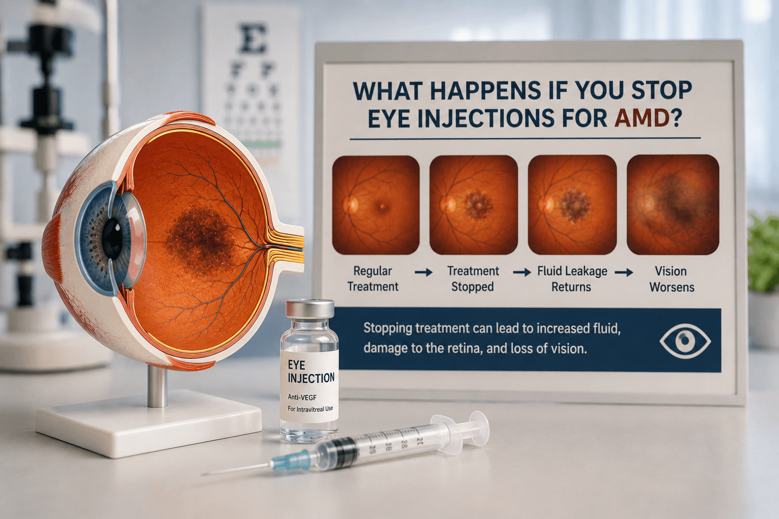
Anti-VEGF Injections
Anti-VEGF agents (ranibizumab, aflibercept, bevacizumab) are the first-line treatment for macular oedema in both CRVO and BRVO. They work by reducing VEGF-driven vascular permeability, decreasing macular fluid. In landmark trials (CRUISE for CRVO, BRAVO for BRVO), ranibizumab produced significant improvements in visual acuity compared to sham injection. Similar results have been demonstrated for aflibercept in the GALILEO and COPERNICUS trials.
Dexamethasone Implant (Ozurdex)
A sustained-release dexamethasone implant (Ozurdex) placed into the vitreous cavity provides anti-inflammatory and anti-oedema effects lasting 3–6 months. It is particularly useful in pseudophakic patients (who have already had cataract surgery), as the primary concern with steroid use — cataract formation — has already been addressed. Ozurdex is also beneficial in patients who have had an incomplete response to anti-VEGF.
Grid Laser
Grid laser photocoagulation to the area of macular oedema was the standard treatment for BRVO-related macular oedema before the anti-VEGF era. It is now used less frequently as a primary therapy, as anti-VEGF produces superior vision outcomes. However, it retains a role as an adjunct or in cases where ongoing injections are not feasible.
Panretinal photocoagulation for ischaemic RVO
Systemic Workup After Retinal Vein Occlusion
A diagnosis of RVO should prompt a systematic review of cardiovascular and haematological risk factors. Recommended investigations include:
- • Blood pressure measurement — essential; often the primary modifiable risk factor.
- • Fasting lipid profile — to detect hyperlipidaemia.
- • Fasting blood glucose and HbA1c — to screen for or monitor diabetes.
- • Full blood examination (FBE) — to detect polycythaemia, thrombocytosis, or anaemia.
- • Coagulation screen — particularly in younger patients or those with bilateral occlusion or recurrent episodes.
- • ESR and inflammatory markers — to exclude vasculitic causes, especially in younger patients.
Referral to a GP or physician for cardiovascular risk factor optimisation is an integral part of RVO management. Controlling blood pressure alone can substantially reduce the risk of a second occlusion.
Sudden vision change in one eye?
Retinal vein occlusion requires prompt assessment and treatment. Book an urgent appointment with Dr Parth Shah.
Central vs Branch Retinal Vein Occlusion
| Central RVO (CRVO) | Branch RVO (BRVO) | |
|---|---|---|
| Area affected | Entire retina — all four quadrants | One quadrant or sector of the retina |
| Severity of vision loss | Often severe; profound central vision loss is common, particularly in ischaemic CRVO | Variable; many patients retain good central vision if the macula is not directly involved |
| Prognosis | More guarded; vision recovery is less predictable, especially in ischaemic cases | Generally better; spontaneous partial recovery is common, and macular oedema responds well to treatment |
| Main complications | Macular oedema, neovascular glaucoma (from iris neovascularisation), vitreous haemorrhage | Macular oedema, retinal neovascularisation (less common than in CRVO), vitreous haemorrhage |
| Treatment | Anti-VEGF injections for macular oedema; panretinal photocoagulation for ischaemic CRVO | Anti-VEGF injections or Ozurdex for macular oedema; grid laser in selected cases; PRP if neovascularisation develops |
Conclusion
Retinal vein occlusion is a sudden, frightening event that can cause significant central vision loss. Anti-VEGF eye injections have transformed management of the most treatable consequence — macular oedema — with large clinical trials demonstrating meaningful vision improvement in the majority of treated patients. Early initiation of treatment, thorough systemic investigation and risk factor control, and regular OCT-guided follow-up are the cornerstones of optimal management.
Frequently Asked Questions
Recovery depends significantly on which type of occlusion occurred, how much of the retina was ischaemic (deprived of oxygen), and how promptly treatment was started. For BRVO with good initial vision and no central macular involvement, spontaneous improvement is common. For CRVO — particularly the ischaemic type — vision recovery is more limited. Anti-VEGF treatment can reduce macular oedema and allow significant vision improvement, but cannot reverse ischaemic damage to retinal nerve cells that has already occurred.

Written by
Dr Parth ShahDr Parth Shah is a subspecialty-trained Canberra ophthalmologist with expertise in cataract surgery, paediatric eye care and strabismus surgery.
Medical disclaimer: This article is for educational purposes only and does not constitute medical advice, diagnosis, or treatment. Always consult a qualified ophthalmologist or healthcare professional regarding any eye condition or before making any healthcare decisions. Read our full disclaimer.
Concerned about your retinal health?
Book a comprehensive retinal assessment with Dr Parth Shah to discuss your vision and treatment options.